60 years old female patient, with neglected leg ulcers, who developed a necrotic cellulitis of the left foot and ankle.

Fig. 1: Initial aspect on admission, showing the extensive necrotic infection of the left foot and ankle, involving also tendon sheaths on the dorsal and plantar aspect of the foot.
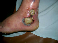
Fig. 2: Lateral view of the left ankle, showing the necrotic infected venous ulcers from which the suppuration has spread in the left foot.
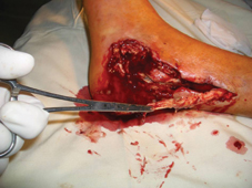
Fig. 3: Surgical debridement.
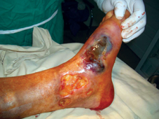
Fig. 4: Oedema and septic blisters of the inner aspect of the left foot and ankle which are usually signs of spreading infection (necrotic cellulitis).
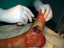
Fig. 5: Thick pus and debris with terrible decay scent flowed out through the incision (a mixture of pseudomonas aeruginosa and streptococcus faecalis). All the necrotic area has to be removed in a life and limb salvage procedure.
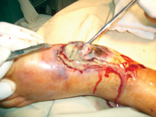
Fig. 6: The area of the septic necrosis is very large, involving all soft tissues of inner and lateral aspect of the left foot and ankle.
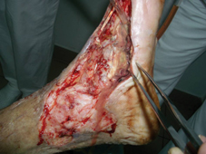
Fig. 7: Even after aggressive surgical debridement, some necrotic tissues still persist, surrounding metatarsal bones and plantar tendon sheaths.
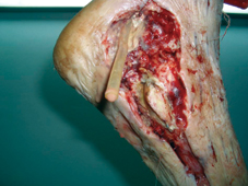
Fig. 8: A plastic tube passive drain has been placed from the lateral aspect to the plantar and inner aspect of the excisional defect. The extern malleolus can be seen in the middle of the lateral excisional area.
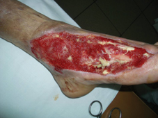
Fig. 9: After about three weeks of passive debridement using LIGASANO® white, the most part of the remaining necrotic tissue has been removed and replaced with good granular tissue.
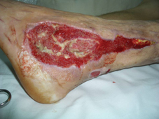
Fig. 10: The same spectacular evolution can be seen on the lateral aspect where the granular tissue covers progressively all the excisional defect, even the external malleolus.
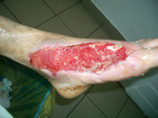
Fig. 11: The same progress of granulation can be seen on the inner aspect of the foot, where still small areas with sloughs and debris persist.
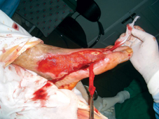
Fig. 12: A wick of LIGASANO® white is inserted from the dorsal to the plantar aspect between the first and the second metatarsal bone, in order to debride all remaining necrotic tissue and to promote a good granulation process.
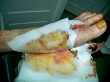
Fig. 13: The LIGASANO® wick is covered by two layers of LIGASANO® white, which are changed 3-4 times weekly, due to the abundant exudate.
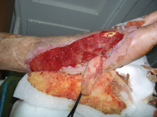
Fig. 14: The passive debridement using LIGASANO® white is very effective, promoting a spectacular granulation as well.
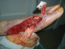
Fig. 15: The LIGASANO® wick is debriding the deep plantar and web spaces, removing sloughs and debris from these areas.
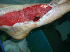
Fig. 16: The granulation tissue progressively covers all the excisional area, even the deep plantar space due to the LIGASANO® wick and sheets of foam.
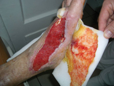
Fig. 17: The spectacular granulation penetrates sometimes the cells of LIGASANO®, producing small unimportant bleeding which stops after dressing removal.

Fig. 18: The LIGASANO® wick is less soaked by the exudate and can be removed once in a week only, as well as the the foam dressing sheets covering the granular area.
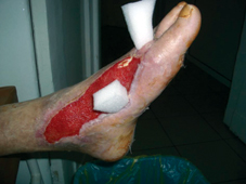
Fig. 19: Finally shorter pieces of LIGASANO® are inserted in the first web space and the plantar space (without crossing between these two spaces), because the new granulation tissue filled the deep tunnel created by sepsis and sloughing. This case is still in process and the granulation area has to be grafted.