Practice of Diabetology Carola Zemlin M.D., Wanzleben/Germany
Anamnesis
50 years old, male, type 2 diabetes mellitus, duration 22 years; ulcer of the right heel since September 1998, diabetic retinopathy, coronar heart disease, state after aortocoronary bypass, wound state 2 according WAGNER, classification ARLT C. Insulin therapy 3x normal insulin; early, noontime and late NPH insulin.
Local pre-therapy
With several ointments, powder and sodium borate, under it progredient deterioration. 67 days of hospital stay because of the ulcer.
Actual findings at 1998/12/17
At the first vistit in our ambulance ulcer of the right heel with a dimension of 20 x 25 mm and a depth of 15 mm, phlogistic reaction of the environmental tissue, smeary coating. Bacterial smear of the wound: streptococcus G1, pseudomonas species. Warm feet, anhidrosis, pulses of the feet poorly palpable, indolent lesion. By reason of the extend findings and the arterial occlusive disease hospitalisation in the surgical clinic, where at the same time the vascular diagnosis takes place. An extensive bilateral arterial occlusive disease exists, no possibility of revascularisation. The amputation of the lower leg as ultima ratio was proposed.
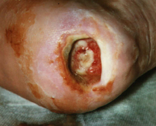
Fig. 1: 1999/02/25
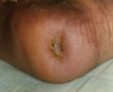
Fig. 2: May 2000
Another visit in our ambulance at 1999/02/25:
Size of the ulcer 18 x 25 mm, depth 5 mm, partly exposed calcaneus. Beginning of the local treatment with sterile LIGASANO® white, soaked with Ringer´s solution and blood of one´s own, daily dressing change. Hypergranulation was cauterised with a AgNO3 pen very carefully. In March 1999 the wound surface reachs skin level and minimises itsself continuously until the final wound closure in May 2000.
Study about the use of LIGASANO® white as initial wound dressing at foot disease caused by diabetics, of Carola Zemlin, M.D., internist/diabetologist. For the evaluation of the study please click here.