49 years old female patient, with type II diabetes treated with insuline and diet. This patient has had a small contusion on her left elbow, from which an extensive sepsis has developed, involving her arm, forearm and hand.
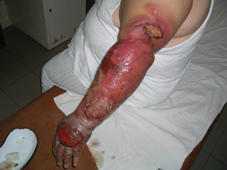
Fig. 1: Initial aspect on admission, with extensive septic epidermolysis and blistering on her left upper limb.
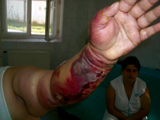
Fig. 2: The volar aspect reveals the circumferential involvement of the left forearm by the septic process.
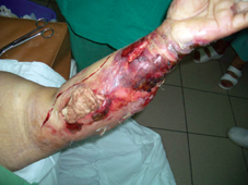
Fig. 3: Surgical debridement began by cutting incisions through the dermis in order to interfere with the lymphatic vessels and stopping the lymph flow and thus the potential septic dissemination of the extensive soft tissue infection.
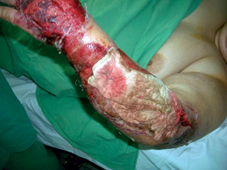
Fig. 4: 24 hours after surgical debridement, when all necrotic tissues and pus have been removed by through-and-through large incisions (from the dorsal to the volar aspect of the left forearm), the remaining defect being packed with gauze pads, soaked in chloramine solution.
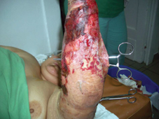
Fig. 5: The haemostatic clamp inserted in the forearm incisional “pocket” shows the large incisions, opening the forearm compartments and leaving a large access to the necrotic areas in order to be debrided.
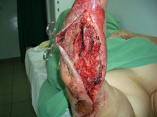
Fig. 6: Detail image of the same case one day after the initial surgical debridement showing the large openings of the forearm compartments.
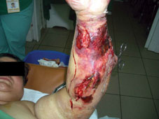
Fig. 7: Volar aspect of the forearm showing the incisions transfixing the skin and underlying tissues in order to delimitate and remove necrotic areas.
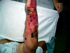
Fig. 8: Four days after initial debridement (septic lesions have been dressed every day with argentic sulphadiazine 5% cream), oedema decreased and necrotic area are well separated from the sorrounding healthy structures.
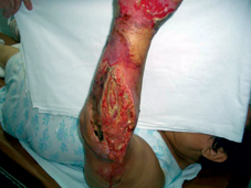
Fig. 9: The most part of the septic discharges are drained off and the necrotic skin is now separated. A small septic discharge still persists from the open dorsal compartment. This is the moment in which the passive debridement with LIGASANO® white has begun.
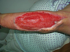
Fig. 10: After about two weeks of passive debridement with LIGASANO® white, a spectacular granulation has been obtained and all debris and septic sloughs have been removed.
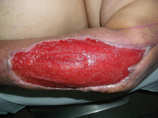
Fig. 11: Detail image of the granulation tissue, that has an excellent quality, with a very good blood flow and little exudate.
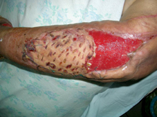
Fig. 12: The granulated area has been grafted with meshed STSG with full take on the distal half of the wound. The proximal half has to be re-grafted, due to a small remaining unseen sinus with septic discharge from the elbow.
This case is still in process.